- This event has passed.
ThermoFisher Seminar

Engineering fluorescence for imaging in cell biology
Michael S. Janes, M.S.
Fluorescence microscopy offers robust, multi-parametric and scalable visualization and analysis of heterogenous populations of cells with spatial and temporal resolution, often critical for effective interrogation in biology. In this seminar, we will discuss a powerful toolbox of probes and applications using fluorescence microscopy and high content analysis. Topics will include:
Practical considerations in fluorescence labeling and detection
Cell structure and organelle dynamics
Cell proliferation, stress, death and internalization
Applications in tumor spheroids and immuno-oncology research
Introduction to spatial biology for higher plex labeling and detection of tissue sections
Beyond conventional: Acoustic focusing at the forefront of advanced flow cytometry
Heaven Le A. Roberts, Ph.D.
As the field of flow cytometry continues to advance, researchers are discovering exciting applications for the growing wealth of information derived from their samples. Innovative reagent solutions, enhanced fluidic and optical engineering, and advanced detection technologies— including imaging integration, spectral unmixing, and high-speed acquisition with robotic automation—have significantly increased the speed, accuracy, and depth of data available. Despite these advancements, challenges remain, particularly in the flow analysis of rare, delicate, and limited samples. This seminar will instruct users in overcoming these challenges using the available technologies in this rapidly expanding field.
Seminar outcomes:
Understand the impact of acoustic focusing technology on flow cytometric analysis
Discover the benefits of a clog-resistant design in maintaining workflow efficiency and tools
to avoid poor quality acquisitions even with difficult samples
Learn how to leverage image-enhanced cytometry data beyond doublet discrimination by
integrating shape, size, intensity, and co-occurrence morphological parameters with
fluorescence detection
Gain practical insights for maximizing data and acquisition efficiency in high-plex
conventional and spectral unmixing experiments
For more information, contact Ryan Gill at ryan.gill2@thermofisher.com
Speakers:
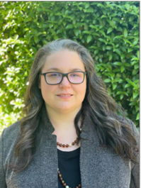
 Heaven Le A. Roberts, Ph.D.
Staff Scientist, R&D
Protein and Cell Analysis
Thermo Fisher Scientific
Eugene, Oregon USA
Heaven’s Ph.D. work at Oregon State University focused on mycotoxin biotransformation and immunotoxicity, which ultimately led to a career in the nutritional specialty products industry. In 2020, Heaven’s experience with cytometric analysis of a wide range of sample types led to her current role developing flow cytometry instrumentation in R&D at Thermo Fisher Scientific. She now develops detection and analysis solutions for customers wishing to expand their understanding of cellular systems using flow cytometry and imaging.
 Michael S. Janes, M.S.
Senior Staff Scientist, R&D, Protein & Cell Analysis
Thermo Fisher Scientific
Eugene, Oregon USA
Mike received his M.S. in 1996 from Bowling Green State University in the United States, where his graduate work focused on mitochondrial biology and comparative biochemistry between parasitic helminths and their mammalian hosts. Mike continued his training as a cell biologist and microscopist at National Jewish Medical Center in Denver, Colorado where he developed fluorescence-based assays for the study of inflammation and apoptosis in respiratory diseases until 2000. He has been a research scientist at Thermo Fisher Scientific developing reagents and assays for fluorescence microscopy since 2000. Mike currently leads the Scientist to Scientist (S2S) Program, intended to connect R&D scientists at Thermo Fisher Scientific with researchers across the globe to drive technical exchange and develop collaborations.

