

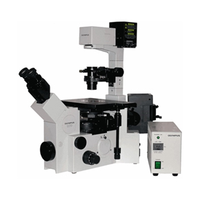
Olympus IX70 Inverted Widefield Epifluorescent Microscope
Basic manually controlled inverted microscope for fluorescent or brightfield samples mounted on slides or in dishes/plates. Mercury metal-halide white light lamp for fluorescence excitation and DAPI, GFP, TRITC filter cubes. An additional Cy5 filter cube may be installed if needed. The microscope is equipped with a QImaging Retiga 4000R monochrome camera for fluorescent pseudo-color or brightfield monochrome imaging. This system also has an optional LCD color module which allows it to capture images of color stained samples in brightfield. Images are acquired using QCapture Pro 7 software.
Objectives: 4x dry, 10x dry, 20x dry LWD, 40x dry and 60x water.
Location: CTAC Imaging Suite (MBI LG-164)
QImaging Retiga 4000R Monochrome Camera with RGB-HM-5 Color Filter
Uses QCapture Pro 7* (Windows 7)
Prior Lumen 200 Mercury Metal Halide Lamp
Objectives
- 4x) UPlanFl NA=0.13 WD=17mm
- 10x) UPlanApo NA=0.40 WD=3.1mm
- 20x) LCPlanFl NA=0.4 WD=6.9mm (Correction Cap)
- 40x) UPlanApo NA=0.85 WD=0.2mm
- 60x Water Immersion) UPLSAPO 60XW NA=1.2 WD=0.28mm
- 60x Oil Immersion) PlanApo NA=1.40 WD=0.15mm (Installed as needed)
- 100x Oil Immersion) UPlanApo NA=0.5-1.35 WD=0.10mm (Installed as needed)
Fluorescent Filter Sets
- Blue / DAPI: Semrock BrightLine DAPI-5060B
- Green / GFP: Semrock BrightLine GFP-3035B
- Red (Short) / TRITC: Semrock BrightLine TRITC-A
- Red (Long) / CY5: Chroma 41008 Cy5 (Installed as needed)
*For online software tutorials, please see the following:
Basic Operation
Capturing Monochrome Images: Standard fluorescent imaging mode, for taking pseudo-color single and multi-channel fluorescence images, or basic B&W brightfield.
Capturing Color Images: For taking true color images of H&E stains, etc.