

STED super resolution imaging is now available to UF researchers
STED super resolution imaging is now available to UF researchers
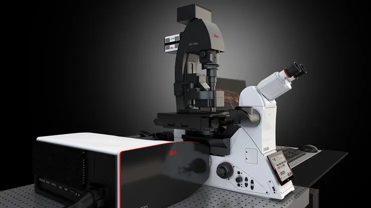
ICBR’s Leica Stellaris 8 laser scanning spectral confocal microscope has been relocated from the CGRC building to its new home in ICBR Cytometry’s Optical Imaging suite in the McKnight Brain Institute (MBI room LG-164). During reassembly, the instrument was upgraded with Leica’s STED super resolution technology. STED (STimulated Emission Depletion) offers the ability to dramatically improve fluorescent image resolution.
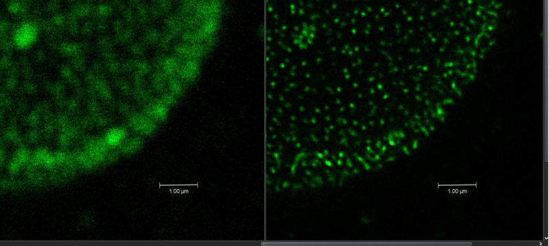
The new STED imaging mode allows for imaging at resolutions down to ~30nm in the XY axes and ~100nm on the Z axis, opening exciting possibilities such as imaging nanoparticles and EVs. STED super-res technology offers rapid confocal point-scanner operation in both 2D and 3D (z-stack) imaging modes, with tissue depth-penetration to potentially 30um.
The example image shown below compares a conventional confocal image (L) captured on our Stellaris 8 with a STED image (R), showcasing the dramatic improvement in resolution. The sample is a cell nucleus with a nuclear membrane pore stain.
Leica Stellaris 8 Highlights:
The system is equipped with a 405nm laser for DAPI/Hoechst imaging, and a tunable wavelength White Light Laser (WLL) which is adjustable from 440 to 790nm for compatibility with a wide range of fluorescent labels.
The Stellaris is also fitted with 5 spectrally adjustable HyD emission detectors (three HyD S, and two HyD X) but may be configured to capture additional channels if scanning by frames or stacks.
Fluorescent Lifetime IMaging (FLIM) technology can help remove autofluorescent background and allows imaging of some closely emitting fluorophores by separating them based on their photon return times. FLIM technology has now been integrated into STED functionality, for improved performance over previous generations of STED.
The Leica Stellaris 8 STED is now operational and available for training.