

Jaw-Dropping Recovery: UF Veterinarians Save Injured Dairy Calf Using Creative 3D Solution
from VetMed
by Sarah Carey
Thanks to a creative solution devised by University of Florida veterinarians using 3D printing technology, a newborn calf that sustained a broken lower jaw soon after birth at the UF dairy farm in Hague has fully recovered and is now an 11-month-old growing heifer in the herd.

Dr. Fiona Maunsell walks with Potato at the UF-IFAS dairy unit in Hague on Dec. 7. (Photo by Jesse Jones)
“We were at the farm soon after she was born, and the staff asked us to look at her because she was standing with her mouth hanging open,” said Fiona Maunsell, BV.Sc., Ph.D., a clinical assistant professor with the UF College of Veterinary Medicine’s food animal reproduction and medicine service. Veterinarians from the service regularly visit the farm, run by UF’s Institute of Food and Agricultural Sciences and located in Hague, Florida, to provide care for its dairy cows and calves.
The day the calf was born, it was seen trying to first stand after birth and nose-diving forward, hitting the ground right on the end of her chin, Maunsell said. Although farm staff initially thought the injury was minor, when the UF team examined the calf more closely, they determined she had a broken lower jaw.

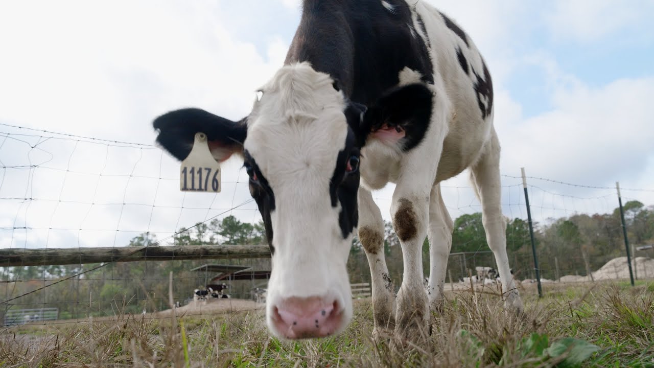
Potato, now nearly a year old, has recovered well from her injury, thanks to treatment by UF veterinarians. (Photo by Jesse Jones)
Maunsell said thousands of calves take their first steps every day, and almost all of them fall down multiple times in the process.
“This is the first time I’ve ever had a calf do this,” she said. “She was very feisty, though, running around her pen. And if you helped hold her mouth closed, she was able to nurse fine from a bottle.”
Although veterinarians are often able to repair breaks in the lower jaw in the field when they are at the front of the jaw, adjacent to or between the front teeth, this calf’s break appeared to be on both sides at the back of the jaw.
“That’s not something we could repair with field anesthesia and surgery,” Maunsell said. She then contacted two of UF’s large animal surgeons to see if she could send the calf for radiographs and an evaluation.

Images from a CT scan of the calf’s bilateral jaw fracture were used to create a 3D printed cast-like device that aided in her healing. At left, the bilateral fractures are clearly seen; the right image shows the complete healing of the bones after treatment. (Photo by Jesse Jones)
The large animal team agreed to see her, and the calf arrived at UF’s Large Animal Hospital as an emergency case on Jan. 7. When radiographs did not shed much light on her injury, the surgical team recommended a CT scan, which involves multiplanar imaging and very effectively showed the bilateral breaks.
“The options for treating a calf with this type of fracture are orthopaedic surgery, which was not economically feasible, conservative management with stabilization of the jaw, or euthanasia,” Maunsell said. “Given that she has had such a great attitude, we thought we’d try conservative management.”
Working with Hongjia He, a graduate student in the Surgical Translation and 3D Printing Research Laboratory run by Adam Biedrzycki, D.V.M., an assistant professor of large animal surgery, large animal surgery resident Heather Roe, D.V.M., sat down and built a jaw “cast” for the calf veterinary technicians named “Potato.”
“We used medical tape to hold her mouth closed to stabilize her jaw, but still allow her to nurse,” said Taralyn McCarrel, an assistant professor of large animal surgery, who worked with Roe on the case. “The tape was challenging to reset with Potato’s feisty nature and started to irritate her skin, whereas the cast placed in a similar fashion was much easier to reset.”